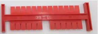
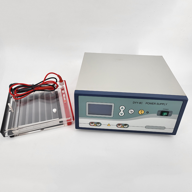
Gel electrophoresis is a routine task in molecular biology laboratories for nucleic acid and protein separation and identification. Scientists use gel imaging systems, also known as gel documentation (gel doc) systems or gel imagers, to visualize bands on DNA and protein gels. Many gel doc systems also capture high-quality images of gels and blots and allow researchers to quantify signal intensity that correlates with protein or nucleic acid amounts in each band.1
To visualize DNA or protein in gels, researchers load their samples with colorimetric- or fluorescence-emitting dyes. Typical DNA gel imagers visualize DNA intercalating dyes, such as ethidium bromide (EtBr), with a harmful ultraviolet (UV) light transilluminator. While these imagers offer excellent illumination, UV light can induce cross-linking and make incisions in DNA strands, affecting sample integrity. Researchers using UV imagers also put themselves at risk for damage to their skin and eyes.1,2 Additionally, EtBr is toxic and mutagenic. As a safer alternative, researchers developed safer fluorescence-based DNA dyes, such as GelRed®, to replace EtBr. Gel Document Imaging System Gel Electrophoresis Equipment

Fluorescent gel imagers efficiently detect fluorescence-based DNA dyes and work with fluorescently-stained protein gels. These imagers can use UV or LED to illuminate fluorescence in gels. LED-based gel imagers are safer compared to UV as they do not damage eyes and skin, and they offer fast and easy DNA and protein visualization. However, LED gel imagers have poor excitation efficiency, which produces a dimmer signal.2-4 These imagers also produce high background noise if their LED light source generates excessive ambient light.
There is an unmet need in gel imaging technology for a system that produces an excellent signal with low background noise and offers robust performance for a variety of fluorescent dyes. Researchers recently optimized laser diodes for gel imaging to improve signal-to-noise ratios. The Gel-Bright™ Laser Diode Gel Illuminator contains 12 laser diodes offering brighter and clearer nucleic acid and protein gel imaging with high sensitivity. This laser illuminator avoids the hazards of UV light while providing performance comparable to UV transilluminators. Unlike blue LED gel imagers, Gel-Bright™ illuminates gels with less ambient light and is compatible with widely used green and red dyes, exciting them more closely to their optimal excitation wavelength. In fact, this laser illuminator is better at imaging green dyes, such as GelGreen® and SYBR® Green, compared to UV-based transilluminators, and red dyes, such as GelRed®, EtBr, and Lumitein™ protein gel stain, compared to blue LED gel illuminators.3
The Gel-Bright™ Laser Diode Gel Illuminator is versatile and user-friendly. It is designed with a lighting angle optimized for even illumination and allows users to adjust the light intensity for ideal signal capture. Due to the device’s design and adjustable filter, it is easy for scientists to access their gels when excising bands during purification protocols.
Overall, this novel laser diode illuminator provides researchers a safe, versatile, and high-performing gel imaging instrument.

Electrophoresis Meaning © 1986–2023 The Scientist. All rights reserved.